Intracranial Pressure Monitoring (measuring pressure on the brain)
Transcript from Video Intracranial Pressure Monitors:
Summary: Someone in a coma from brain injury or bleed may have high intracranial pressure
(ICP). ICP is measured via a monitor, one of two types: (1) non-draining monitor placed in brain tissue; or (2) External Ventricular Drain (EVD) placed within the brain's lateral ventricle (fluid-filled chambers within the brain that we all should have) to drain. Different ICP monitor and EVD brands exist, secured to the head using a screwed in bolt or a stitch. A varying size hole can be drilled bedside using cranial access kits to measure pressure, temperature, oxygen, and more. The advantage of draining fluid is that it reduces pressure, with safety and efficiency of care being keys to recovery.
Intracranial Pressure Monitoring: Understanding ICP Monitors, EVDs and Cranial Access
When a person experiences a severe traumatic brain injury (e.g. from a car accident), bleed within or around the brain (intracranial hemorrhage), fluid pressure build-up due to obstruction of normal flow channels (hydrocephalus), or another neurological condition (e.g. brain swelling from inflammation or infection) that places the brain at risk, one of the most important measurements for clinicians to effect care is monitoring intracranial pressure (ICP).
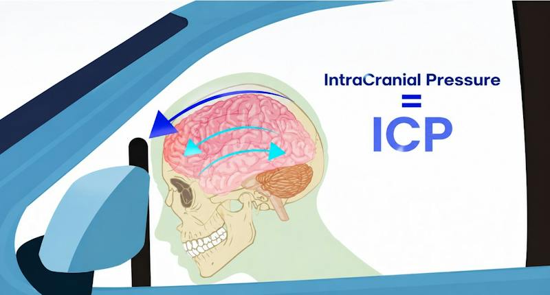
After age 2 (when the soft spots or fontanelles of the baby close completely), the skull is typically a hard, enclosed space (like a hard coconut) containing 3 main items: brain, blood (typically inside blood vessels), and clear cerebrospinal fluid (CSF) bathing the brain. Increased pressure on the brain can result when any of these three items increases in size – like from brain swelling, blood vessel engorgement increasing blood flow, or CSF flow pathway blockage (hydrocephalus). Increasing brain or fluid volume can result in reduced blood flow and “secondary injury” from stroke-like effects, or simply cause direct damage to the brain by compressing it. So knowing the pressure, ensuring it is not too high, and correcting it if it is, can offer life-or-death treatment options.
That’s why ICP monitoring plays such an important role in neurocritical care. The Brain Trauma Foundation includes ICP monitoring in its guidelines for managing appropriate patients with severe traumatic brain injury, where changes in pressure can help guide critical treatment decisions. Many institutions and programs specifically deal with intracranial pressure management, worldwide.
But before an ICP monitor can measure anything, clinicians first need something even more fundamental: safe, controlled access through the skull.
That is where cranial access systems become an important part of the monitoring process.
What Is Intracranial Pressure?
Intracranial pressure refers to the pressure inside the skull.
Under normal circumstances, the brain, blood supply, and cerebrospinal fluid exist within a carefully balanced environment. Trauma, bleeding, swelling, obstruction of CSF (cerebrospinal fluid) circulation, or other neurological conditions can disrupt that balance and cause ICP to rise.

The most accurate indicator of current status is the patient’s neurological exam, when unaffected by sedative medications or other effects (like alcohol, drugs, etc.). A “coma” is a condition of being essentially unconscious but alive, assessed by reviewing eye opening, verbal response, and movement response to command or painful stimulus. Typically, anyone in a coma must receive intracranial pressure monitoring, per the existing standard of medical care – hence the need for ICP monitoring, which can exceed 50,000 cases per year in the United States alone.
ICP monitoring provides continuous physiologic information that can help the care team understand what is happening inside the skull and guide treatment decisions alongside imaging, neurological examination, and other clinical information.
How Is Intracranial Pressure Measured?
There are a few different ways physicians can monitor pressure inside the skull, but two main categories of approaches are used most often:
(1) Intracranial Pressure Monitor (ICP monitor) placed into the substance (not fluid-filled chamber or ventricle) of the brain – placed using sterile conditions (cleaning solution), and drilling a hole, typically a tiny bolt is screwed into the skull, with a fiberoptic or similar probe placed into the brain itself. The advantage of this system is that it does not go through much brain (hence reducing risk of brain damage or hitting a blood vessel inadvertently resulting in bleed). The disadvantage is that since this is in the substance of the brain, but not the fluid-filled ventricle, if the ICP is high, then draining fluid from the ventricle is not an option (as it would be in the case of an external ventricular drain, or EVD). Typically the period of swelling may last many days, but then subside, so the ICP monitor can be removed after consistent days of the ICP remaining normal. Of note, other parameters than pressure (like oxygen levels, electrical activity, or blood flow locally) can be measured by similar probes placed alongside or separately, and treatment plans may be adjusted according to such acquired data – while much of this is newer or experimental, with paradigms being established for treatment based upon them;
(2) External Ventricular Drain (EVD) placement is done using specified landmarks, with incision and hole drilled usually just behind the forehead along the middle of the eye (mid-pupillary line), after marking, cleaning, and numbing the scalp; the procedure is usually done on the right side of the brain (since most people are right-handed, and the left brain typically controls the right side of the body as well as speech, so if a bleed did occur (as it may occur in about 5% of cases) and resulted in brain damage, a right-handed person would not be as affected in writing or speech. The drain is advanced through the brain until it reaches clear fluid in the ventricle (about 50-mm inside), and kept in place, to drain this clear fluid hence reducing (and continuing to monitor) intracranial pressure. Newer modalities combine both fiberoptic-type properties and drainage simultaneously, whereas older drains allowed either measurement or drainage at a given time, so when you measured the ICP you weren’t able to relieve it by draining, and when you drained you didn’t know what it measured. Technically, an EVD does both – drains and monitors ICP – but since conventionally the ICP monitor term referred to non-EVD devices, the term stuck that ICP monitors refer to intraparenchymal monitors (in substance of brain), so hopefully that doesn’t cause too much confusion.
What is Cerebrospinal Fluid (CSF)?
Cerebrospinal fluid is a clear fluid that is an “ultrafiltrate” or a filtered product of blood, present in all of us. It normally circulates around the brain and spinal cord, helping cushion and protect them before being reabsorbed by the body. Problems can occur when that normal circulation or absorption is disrupted. This can happen following a brain bleed, traumatic brain injury, swelling, infection, or with a condition known as hydrocephalus.
Hydrocephalus can be thought of as a plumbing backup inside the brain. CSF continues to be produced, but if its normal pathway becomes blocked or the body cannot absorb it properly, fluid can begin to accumulate within the ventricles. Because the skull is a fixed space, that buildup can contribute to increasing intracranial pressure.
An EVD temporarily creates another pathway for that fluid to leave. Clinicians can carefully control how much CSF is drained while simultaneously monitoring ICP, allowing them to respond to changes in pressure as part of the patient's overall neurological care.
So, although an ICP monitor and an EVD can both provide continuous information about intracranial pressure, they serve different purposes. An IPM is primarily a monitoring device. An EVD provides both monitoring and the ability to therapeutically drain CSF when indicated.
For clinicians, that distinction is important when determining the appropriate monitoring strategy based on the patient's condition, imaging, anatomy, and treatment needs. But whichever system is selected, both begin with the same essential first step: safe, controlled access through the skull.
ICP Monitor vs. EVD: What Is the Difference?
The simplest distinction is that an ICP (intraparenchymal) monitor can be used primarily for monitoring, while an EVD can provide both monitoring and drainage.
An intraparenchymal pressure sensor is inserted through a small opening in the skull and positioned within brain tissue. Once connected to its monitoring system, it can continuously report intracranial pressure.
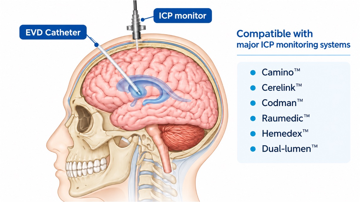
An EVD goes a little farther. Instead of stopping within the brain tissue, the catheter is carefully guided into one of the brain’s lateral ventricles, which are fluid-filled spaces inside the brain (see figure below, excerpted from the video on Intracranial Pressure Monitors at https://www.phasorhealth.com/enlighten/) – highlighting positional differences between ICP monitor vs. EVD, and some common current types of ICP monitors.
Think of the ventricles like small reservoirs that hold cerebrospinal fluid (CSF). Once the catheter reaches that space, it is connected to an external system beside the patient’s bed. That system allows the care team to continuously monitor pressure and, when needed, drain CSF in a controlled way to help relieve pressure inside the skull.
When appropriate, CSF can be drained through the EVD to help control elevated intracranial pressure. Because of this ability to both monitor and intervene, EVD placement is a fundamental procedure throughout neurocritical care.
The appropriate approach depends on the patient's condition, anatomy, clinical objective, physician judgment, and the monitoring technology available at the hospital.
Cranial Access Is the First Step
Whether the physician is placing an EVD or an intraparenchymal monitor, the device must first pass through the skull.
That typically requires creation of a small burr hole or twist-drill opening.
The size and depth of the opening in the skull matter because ICP monitoring systems are not all designed the same way. Different manufacturers may use different bolts, catheters, probes, introducers, and methods for securing the device in place.
Each ICP monitor system that is secured to the skull using a “bolt” needs a very specific size hole, at fractions of millimeters. These may include 2.70-mm, 4.50-mm, or 5.30-mm, or 6.35-mm size holes, for example. Major ICP monitoring systems need specified sizes.
Different ICP Monitors Can Require Different Burr-Hole Sizes
This is an easily overlooked part of ICP monitoring. Hospitals may use one monitoring system in the ICU, another for brain-tissue oxygen monitoring, and another for particular neurosurgeons or patient populations. Those systems can require different cranial-access dimensions.
That means a hospital's cranial access strategy should ideally work with the monitoring technology clinicians already use.
An example of a comprehensive Cranial Access Kit, uniquely customizable to the needs of a hospital to a great extent, is the Phasor Cranial Access Kit – allowing choices of drill drivers (electric or manual), drill bit size(s) (from 2.70-mm to 6.50-mm), and over 30 different items including cleaning solution, instruments for incising, manipulating, or suturing scalp, and dressings, for all-in-one solutions of access kits, varying per need and region. The multiple drill diameters and lengths designed to support a broad range of ICP monitoring and drainage systems. These kits are provided with all the instruments and items needed to typically perform procedures bedside (as they are often done emergently, as “time is brain” – in the Intensive Care Unit or ICU, or Emergency Room or ER, much more frequently than the Operating Room (OR) in adults.
Current Phasor configurations support commonly used technologies, specifically drill bit sizes, including those compatible with Camino™, Cerelink™, Codman™, Hemedex™, IRRAS™, Licox™, Raumedic™, and other intracranial pressure monitoring or drainage systems (with respective company trademarks noted). Each Phasor type of drill driver (electric or manual) offers a quick-connect capability, allowing any of the sizes noted above to be fit, and customized in about 3 seconds to connect. Phasor currently offers a range of drill-bit diameters and lengths, along with interchangeable multi-bit configurations designed to support practically every major ICP monitoring system and a wide range of cranial access needs.
By offering different neurosurgery caregivers (neurosurgeons, neurosurgery residents, or advanced practitioners such as physician assistants or nurse practitioners) their own option of drivers (electric or manual Legacy driver), varying drill bits, and drug-containing vs. drug-free kits, the customizability to individual ICP monitors also exists.
Rather than requiring hospitals to build completely separate cranial access setups around every monitoring platform, this allows cranial access to be standardized while still maintaining compatibility with the devices clinicians prefer.
Why Drilling Control Matters
Creating a burr hole may represent only one portion of an ICP or EVD procedure, but it occurs immediately adjacent to the dura and underlying brain.
Controlled depth is therefore important.
One of the biggest concerns during cranial drilling is something called “plunging.” This happens when the drill passes through the skull and continues farther than intended instead of stopping at the right depth.
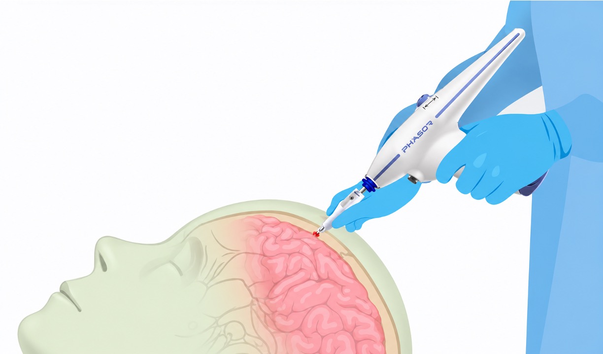
That is why a cranial access drill should be evaluated as more than simply a tool for creating a burr hole. What happens at the moment the drill breaks through the skull matters. Depth control, drill design, and the presence of a physical safeguard can all play an important role in limiting unintended forward movement.
Phasor’s powered cranial drill uses a reinforced mechanical anti-plunge stopper that physically limits how far the drill can advance rather than relying only on the drill to stop rotating. Tested to withstand 50 pounds of downward force, the stopper also helps support a stable, perpendicular approach to the skull. The concern of so-called “self-stopping drills” is that while they may stop rotating (after sensing beyond the skull’s loss of resistance), the drill bit itself can be pushed into the brain and “plunge” – as has been described in published literature at an incidence up to 1 in 200 (Vogel et al, 2011).
Why does perpendicular matter? Drilling straight through the skull helps create a more controlled burr hole and reduces the chance of the drill traveling at an unintended angle. With traditional cranial drills, surgeons generally maintain that position through visual alignment, hand control, tactile feedback, and experience. Phasor’s adjustable stopper (set by gauging a specific patient’s skull thickness) provides an additional point of flat contact against the skull, helping support both angle control and depth control during drilling.
For experienced neurosurgeons and residents alike, that added mechanical safeguard can provide another layer of control while complementing proper technique, training, supervision, and clinical judgment. With now over 32,000 consecutive procedures without plunging with proper drill use, the Phasor Drill Stop Extension has an unparalleled anti-plunging statistic vs. any other conventional or non-conventional technique.
The Future: One for All in ICP monitoring
ICP monitoring technology continues to evolve, but every invasive system begins with the same essential requirement: controlled access through the skull.
A cranial access platform designed around multiple drill sizes and configurations can help hospitals accommodate the range of EVD, ICP, and advanced neurological monitoring technologies used across their neurosurgery and neurocritical care teams.
Phasor Health's Cranial Access Kits are designed around that flexibility, with fixed and interchangeable drill configurations, multiple diameters and lengths, sterile single-use options, mechanical anti-plunge protection, and compatibility with leading intracranial monitoring systems.
For hospitals, that creates an opportunity to standardize the cranial access process without eliminating surgeon choice or changing the ICP technology already in use.
Watch: Intracranial Pressure Monitors
Learn more: Explore Phasor Cranial Access Kits